


Varicose veins are pathological expansion of veins located on the surface, which are characterized by increased diameter and length, resulting in cylindrical, serpentine, cystic, and mixed changes in the trunk of the vein. Today, varicose veins are a common pathology, and women are nearly three times more likely to get sick than men. This is mainly due to the anatomical characteristics of the body and certain loads on the lower limbs during pregnancy.
Usually, varicose veins are primary and secondary. In the first variant, the disease is caused by the initial weakness of the wall of the large vein, which is a congenital dysfunction of the wall of the large vein located under the skin or the valve. The development of secondary venous disease is affected by deep vein thrombosis or acquired valvular insufficiency caused by pregnancy, heavy physical exertion, and prolonged standing.
As the hydrostatic pressure in the veins increases, the diameter of these blood vessels expands and the impaired function of the valves worsens. All this interferes with the blood circulation of the superficial veins. Due to the insufficient function of the peripheral veins, blood flows back from the deep veins to the great saphenous vein. The great saphenous vein is overstretched and begins to peristalize, forming various forms of expansion. In the future, due to obvious stagnation, tissue malnutrition will result in ulcers, eczema and dermatitis.
Varicose veins of the lower extremities
This disease is characterized by the formation of cystic dilatation, serpentine tortuosity, increased length, and valve insufficiency in the vein walls.
Generally, varicose veins of the lower extremities occur in 20% of the population. In addition, before puberty, it also affects boys and girls. But compared with men, adult women are more susceptible to varicose veins. In addition, the number of patients increases with age. This can be explained by the reorganization of the hormonal background in the female body caused by pregnancy and menstruation, which can lead to weakened venous tone, dilation, traffic and saphenous vein valve insufficiency, arteriovenous shunt and venous circulation disorders.
So far, the real cause of the development of varicose veins of the lower extremities is still unknown. It is speculated that insufficient valve function and increased venous pressure are related to the cause of disease development. Considering all the factors that lead to the occurrence of pathological processes in the veins of the lower extremities, there are two types of varicose vein diseases: primary and secondary.
Primary varicose veins on the surface are characterized by the presence of normal deep veins. In the case of secondary varicose veins, various complications such as deep veins, arteriovenous fistulas, congenital venous valve loss or hypoplasia play an important role.
Risk factors related to the formation of varicose veins in the lower extremities include: increased hydrostatic pressure in the trunk of the veins, thinning of the vein walls, impaired smooth muscle cell metabolism, and blood flow from deep veins to superficial veins. This reverse movement of blood in the form of vertical and horizontal return leads to the gradual expansion, lengthening and bending of nodules in the subcutaneous, that is, superficial veins. The last link of the disease is represented by cellulitis, dermatitis and vegetative venous ulcer of the calf.
Symptoms of varicose veins in the lower extremities include complaints from patients about existing dilated veins, which can cause cosmetic inconvenience, with a certain degree of severity, and in some cases, pain in the lower extremities, night cramps, and nutritional changes in the legs.
The expansion of venous blood vessels can be clearly seen in the patient's upright position from tiny "star-shaped" and mesh nodes to roughly peristaltic torso, as well as nodes and clusters. Nearly 80% are surface lesions of the main trunk and branches of the great vein, and 10% are in the small saphenous vein. In addition, in 9% of patients, there is bilateral venous disease during the pathological process.
As a result of the gradual process, the patient begins to fatigue rapidly, with a certain degree of severe and swelling of the legs, cramps in the calf muscles, swelling of the legs and feet, and paresthesias. In addition, the legs are mostly swollen in the late afternoon, but the swelling disappears after sleep.
In many cases, varicose veins will be accompanied by acute thrombophlebitis on the surface of the vein, which is manifested as redness, cord-like, and painful venous compaction, which is characterized by dilation and periphlebitis. Varicose veins often rupture due to minor injuries, which can cause bleeding. Normally, the blood from the ruptured node flows in a fluid state, and the patient sometimes loses a considerable amount of blood.
In addition, there is no certain difficulty in the diagnosis of varicose veins of the lower extremities and the addition of CVI based on the patient's main complaint, medical history, and objective examination results.
A basic value of diagnosis is the ability to determine the valve status of the main and communicating veins, and to assess the patency of the deep veins.
Causes of varicose veins
This pathological process is characterized by dilation of the veins located on the subcutaneous surface, and is related to the insufficient function of the valves in the veins and impaired blood circulation. Varicose veins are one of the most common vascular diseases in half of the working-age population.
Generally, there are several factors that induce disease that will lead to the development and progression of the disease. It has not been confirmed that genetics have a clear contribution to the appearance of varicose veins. The appearance of this pathological process may currently be affected by conditions caused by changes in the nature of diet, lifestyle and hormonal background.
In addition, the occurrence of this pathological process is related to the wrong organization of the work process. Many people spend a lot of time standing or sitting depending on their work, which has a considerable adverse effect on the valve devices of the veins of the lower limbs. In addition, work related to heavy physical labor is considered disadvantageous, especially in the form of heavy loads on the legs when lifting weights.
Today, long-term travel or flying can cause blood stasis in the legs and is a risk factor for the formation of venous disease, which negatively affects the blood flow system in the veins. In addition, wearing tight underwear will compress the veins in the groin area, and a corset will increase the pressure in the peritoneum, so it is not recommended to wear it all the time. This also applies to high heels with uncomfortable insteps.
Recurrent pregnancy is a proven risk factor for varicose veins. This can be explained as the enlarged uterus increases the pressure in the peritoneum, and progesterone destroys the source of elastic fibers and collagen contained in the vein wall. In addition, diseases such as rheumatoid arthritis, osteoporosis, and changes in hormonal status can also increase the risk of this pathological process.
The typical cause of varicose veins is the particularity of the structure of the lower extremities. There is a superficial venous system, the great saphenous vein, the deep venous system of the thigh and calf, and the perforating vein that connects the first two systems. In the case of normal blood circulation, blood flow in the lower limbs occurs in 90% of the deep veins and 10% of the superficial veins. But in order to allow blood to flow to the heart, not the other way around, the valves on the vein walls close violently, not allowing blood to pass from top to bottom under the influence of gravity. Muscle contraction is also important and contributes to normal blood flow. In addition, in the upright position, blood stagnation develops and venous pressure begins to increase, which causes them to expand. In the future, insufficient function of the valve will be formed, which will cause the valve leaflets to not close, resulting in incorrect blood movement from the heart.
The valves of the deep veins are affected particularly quickly due to the maximum load. And in order to reduce excessive pressure with the help of the perforating venous system, blood flows into the veins located under the skin, which are not designed for large amounts of blood. All of these can lead to excessive extension of the vein wall, which results in the formation of characteristic varicose veins. However, more and more blood continues to flow into the deep veins, resulting in perforating venous valve insufficiency. There is no certain obstacle to the blood flow in the horizontal position. It first enters the deep vessels and then into the superficial vessels. Finally, CVI will show signs of edema, pain, and trophic ulcers.
Symptoms of varicose veins
Varicose veins are characterized by dilation of the veins located under the skin, showing cystic or cylindrical changes. With this pathological disease, coiled veins appear on the skin surface of the legs and feet. The greatest appearance of varicose veins is formed after prolonged or heavy physical exertion. During or after pregnancy, vein dilation is common in young women.
The early characteristic of varicose veins is that there are few non-specific symptoms. At this time, the patient will quickly feel tired, and the legs will continue to be heavy, burning, and bursting, especially after physical exertion. In addition, sometimes there will be transient edema and pain along the entire length of the vein. At the same time, in the late afternoon, after prolonged static load, the ankle and back of the foot swelled. Some features of edema are that they disappear after a night's rest in the morning. At this stage, there are usually no obvious signs of varicose veins. However, these initial symptoms should be a signal for the patient to see a specialist to prevent the progression of varicose veins.
This disease is characterized by slow development, sometimes as long as several decades. Therefore, due to improper treatment, varicose veins progress to form CVI (chronic venous insufficiency).
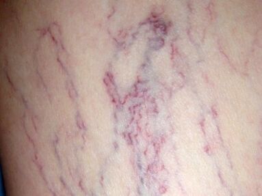
An important symptom of the disease is also spider veins, which are spider webs made up of slightly dilated capillaries that are actually visible under the skin. Sometimes, eliminating hormonal imbalances, eliminating saunas, and sunbathing can make you forget such diseases as varicose veins once and for all. But basically, these spider veins are the only signs that the superficial veins overflow and form varicose veins. Therefore, the appearance of even trivial signs of this kind should be used as a signal to consult a surgeon.
In addition, varicose veins represent cosmetic discomfort. Therefore, in order to solve such problems, doctors will perform surgical operations.
Varicose vein degree
This disease can show varying degrees of severity and is characterized by different structures, which are related to its clinical symptoms. Generally, there are several types of structures for veins that expand on the surface. The first type, the main type, is characterized by the expansion of the main saphenous vein, but no tributaries are added. The second type, or loose, is an extension of a similar network with many branches. Varicose veins of this type are detected at the very beginning of the disease. But for mixed types, there will be a combination of the first two types, and the third type is more common than the others.
The symptoms of varicose veins are directly proportional to the stages of the pathological process, and are divided into compensatory phase, subcompensation phase and decompensation phase.
In addition, the ICD of varicose veins distinguishes pathology from ulcers, inflammations, coexisting ulcers and inflammation of the lower limbs, and varicose veins without inflammation or ulcers.
The first-degree varicose veins are characterized by moderate and significant expansion of the veins on the surface along the trunk or branches, without certain manifestations and traffic characteristics of the surface venous valve insufficiency. The patient has mild leg pain, with a certain severity, fatigue under the background of prolonged fatigue. Diagnostic tests performed showed satisfactory valve function, and slight swelling of the veins under the skin indicated poor venous outflow function of the affected limb. The first degree VL corresponds to the compensatory phase of varicose veins.
The second-degree varicose veins are characterized by dilated superficial veins, and according to functional tests, the valve fails. In the process of impaired venous outflow, the lymphatic system of the limbs is insufficiency, manifested as edema of the feet and legs. Characteristic swelling of the lower limbs will appear after prolonged exertion, and the swelling will disappear after horizontal rest. In addition, there is persistent severe pain in the affected limb. The second degree of disease is characterized by the correspondence of sub-compensatory stages.
Third degree varicose veins, dilated superficial veins, deep vein valve dysfunction, perforation and saphenous veins, leading to persistent venous hypertension in the distal limbs. This is the cause of microcirculation disorders and the formation of trophic ulcers. At the same time, skin pigmentation appears in the calf area, which initially manifests as a rigid pathological process. But the feet and legs, especially if there are nutritional disorders, are characterized by persistent swelling. This is related to blood outflow disorder, organic limb lymphatic system damage and secondary lymphatic stagnation. The symptoms of third-degree varicose veins are very obvious, varied and constant.
With the further development of varicose veins, the area of trophic ulcers has expanded, and dermatitis and eczema have appeared, indicating that the condition has entered the fourth stage. The last two severity levels represent the decompensation stage of the pathological process. In this case, not only local but also general hemodynamics are disturbed. Impaired myocardial contractility can be detected using ballistic cardiography, and 80% of patients with decompensated varicose veins can detect impaired myocardial contractility.
An important point in choosing the right treatment is to determine the degree of varicose veins and the type of dilated superficial veins.
Varicose Vein Treatment
The comprehensive treatment of leg varicose veins is considered to be a complex process, which is proportional to the severity of the disease. Usually, surgery and conservative treatment methods are used.
Varicose veins can be treated without surgery, and only in the initial stages of the pathological process will produce positive results, at which time the appearance on the skin is slightly manifested, which moderately reduces the ability to work. As a conservative treatment, this treatment is also used due to contraindications to surgical intervention. In addition, in order to prevent the recurrence of varicose veins, this method must be used during the postoperative period.
During conservative treatment, the severity of risk factors can be reduced by using sufficient physical activity, elastic compression, drug therapy, and physical therapy. Only the combination of all these treatment measures can guarantee positive results.
First, they identified the risk factors for varicose veins and tried to influence them. In addition, even if there are no symptoms of varicose veins, people with certain risk factors and genetic predispositions need to consult a phlebologist twice a year for ultrasound examination of the veins. Lower limbs. In addition, if there are no complications such as thrombophlebitis or thrombosis, regular training of the veins of the lower limbs is recommended. This includes walking more, wearing only comfortable shoes, swimming, biking, and jogging. All physical activities should be performed using elastic compression. Sports with injuries to the lower extremities are absolutely taboo, and sports such as mountain skiing, tennis, volleyball, basketball, football, and various martial arts that have a heavy load on the veins of the lower extremities must also be excluded. As an exercise related to weightlifting.
At home, they perform simple exercises on the advice of experts. As a general rule, the legs should be in an elevated position for a few minutes before starting to exercise in order to prepare the body for certain types of exercise. The speed of exercise and the choice of speed are strictly and individually selected for each patient, taking into account his physical ability. But the main content of this kind of physical education is its regularity. In addition, it is recommended to use a contrast shower every day and massage the legs alternately with warm and cold water for five minutes.
Elastic compression is a method of using bandages or compression stockings to treat varicose veins. In this case, muscle compression occurs in a dosed manner, thereby improving blood flow through the venous vessels and preventing stagnation. Due to the artificial maintenance of blood vessel tension, the veins stop dilating, thereby preventing thrombosis.
For the treatment of various stages of varicose veins, intravenous drugs are used to gradually strengthen the vein wall. All medications for varicose veins can only be prescribed by the attending physician, so self-administration is not recommended. However, topical treatment in the form of ointments and gels without signs of thrombophlebitis or thrombosis is not advisable.
In physical therapy methods, lasers, electrophoresis, magnetic fields, and the use of reverse kinetic currents have the best results.
Varicose veins are surgical diseases that can be completely cured after surgery. Generally, there are several types of surgical treatments (phlebectomy, sclerotherapy, and laser coagulation), which directly depend on the severity of the pathological process and its location.
During phlebectomy, varicose veins are removed. The main purpose of surgery is to eliminate pathological blood discharge by removing the main trunk of small or large superficial veins and ligating the perforating veins. However, this kind of surgery cannot be performed with diseases that only worsen the existing condition; late varicose veins; pregnancy; existing purulent processes and old age. Phlebectomy is performed using endoscopic treatments, which makes this operation unsafe.
During hardening, sclerosing agent is injected into the dilated venous blood vessels, which causes the walls of the vein to join, thereby stopping the blood flow through it. As a result, the pathological outflow of blood is stopped, and cosmetic defects are eliminated at the same time, because the veins are now collapsed and are almost invisible. However, sclerotherapy is only effective when the main branch is enlarged, so the scope of application is limited. The advantage of this kind of surgical intervention is that there is no scar after the operation, the patient does not need to be hospitalized, and in the post-hardening period, the patient does not need special rehabilitation treatment.
Laser coagulation is based on the destruction of the vein wall by its thermal effect. Due to this process, the venous cavity is sealed. This surgical method is only suitable for veins that are enlarged to 10 mm.
Prevent varicose veins
The prevention of this disease can be primary, it can prevent the development of varicose veins and secondary-in the presence of pathological processes.
At present, most people attach great importance to the prevention of this disease. Simple measures on a regular basis can significantly reduce the occurrence and further development of varicose veins. In this case, it is very important to exercise more first, and to alternate swimming, running, walking, and cycling under a long static load. You should also perform simple exercises in the workplace.
With existing varicose veins, you must try to raise your legs as much as possible. Fight against excess weight and prevent it from gaining. It is also important to walk in comfortable shoes, with a maximum heel height of no more than five centimeters, and use orthopedic insoles when necessary. In addition, during pregnancy, when taking estrogen or oral contraceptives, an ultrasound scan must be used to examine the veins of the lower extremities.